Diagnostic imaging
Radiology
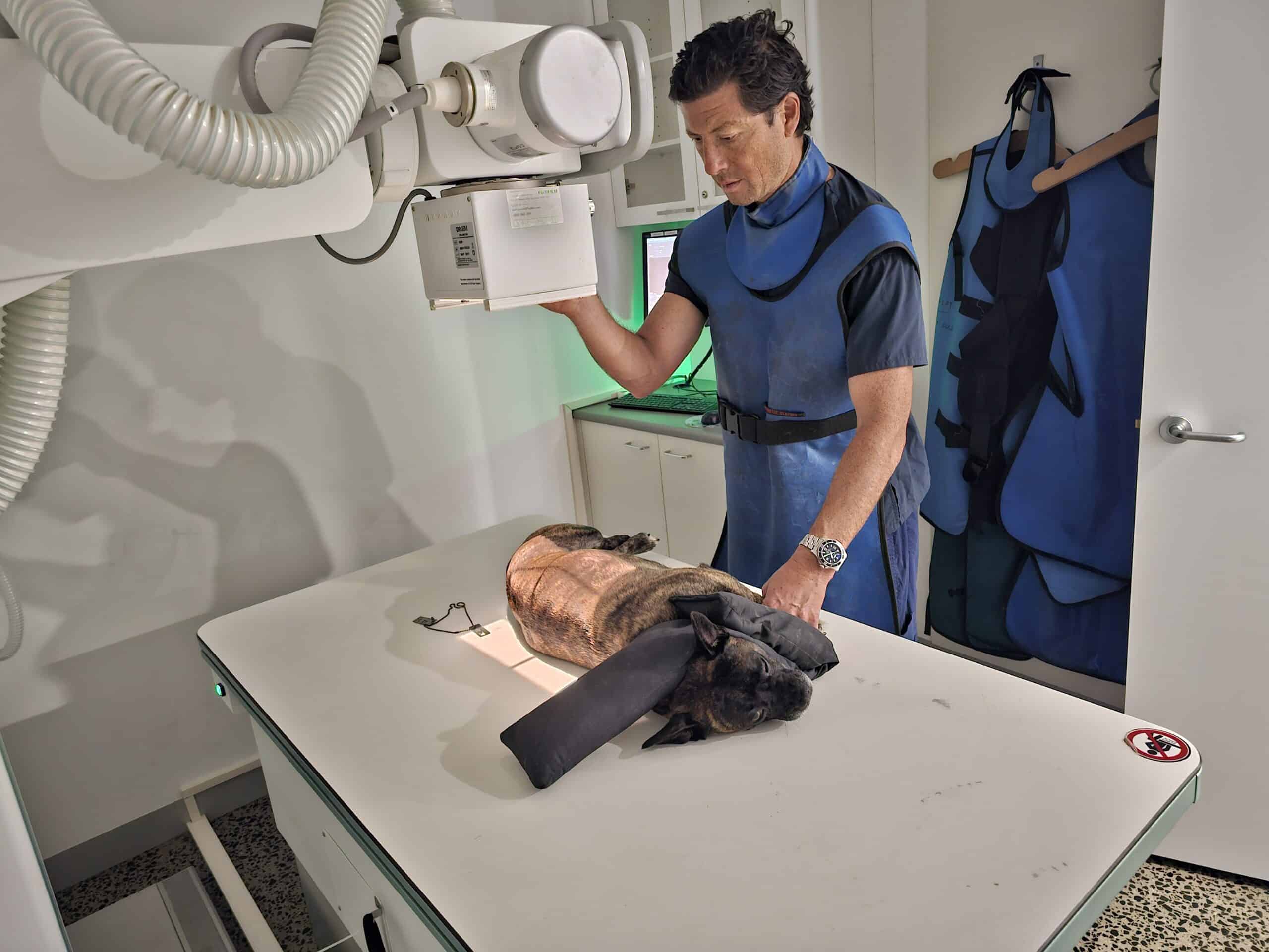
Radiology plays a crucial role in veterinary orthopedics by providing detailed images that help diagnose and manage musculoskeletal issues. Here’s an overview of its significance:
Types of Radiological Imaging
X-rays: Helpful in diagnosing fractures, joint dysplasia, tumors, and arthritis.
The most common imaging modality used to assess bone structure and joint integrity.
Ultrasound: Useful for evaluating soft tissue structures, such as muscles, tendons, and ligaments.
Can help identify fluid accumulation and soft tissue injuries.
Computed Tomography (CT): Provides cross-sectional images, offering detailed views of complex bone structures. Useful for pre-surgical planning and assessing certain types of fractures or lesions.
Magnetic Resonance Imaging (MRI): Excellent for visualizing soft tissues, including ligaments, cartilage, and the spinal cord. Often used for more complex cases, such as intervertebral disc disease or soft tissue tumors.
- Applications in Orthopedics
Fracture Diagnosis: Identifying the type, location, and severity of fractures.
Joint Evaluation: Assessing conditions like hip dysplasia, elbow dysplasia, and patellar luxation.
Tumor Identification: Detecting bone tumors and evaluating their extent.
Post-Surgical Assessment: Monitoring healing after orthopedic surgeries.
Benefits of Radiology
Non-Invasive: Most imaging techniques are non-invasive and provide crucial information without requiring exploratory surgery.
Guiding Treatment: Helps veterinarians create targeted treatment plans, whether surgical or conservative.
Monitoring Progress: Allows for the tracking of healing and recovery over time.
Conclusion
Radiology is an essential tool in veterinary orthopedics, enhancing diagnostic accuracy and treatment outcomes. Veterinarians often collaborate with radiologists to interpret images and ensure comprehensive care for musculoskeletal conditions.
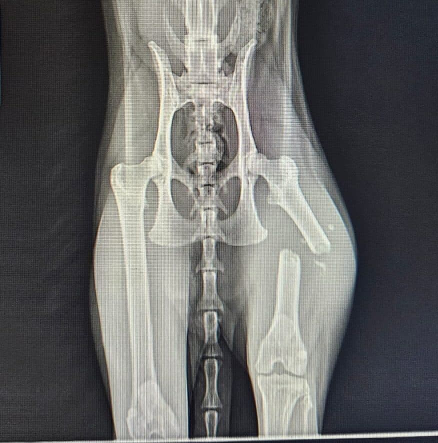
Transverse midshaft femoral fracture in a cat
CT Scan
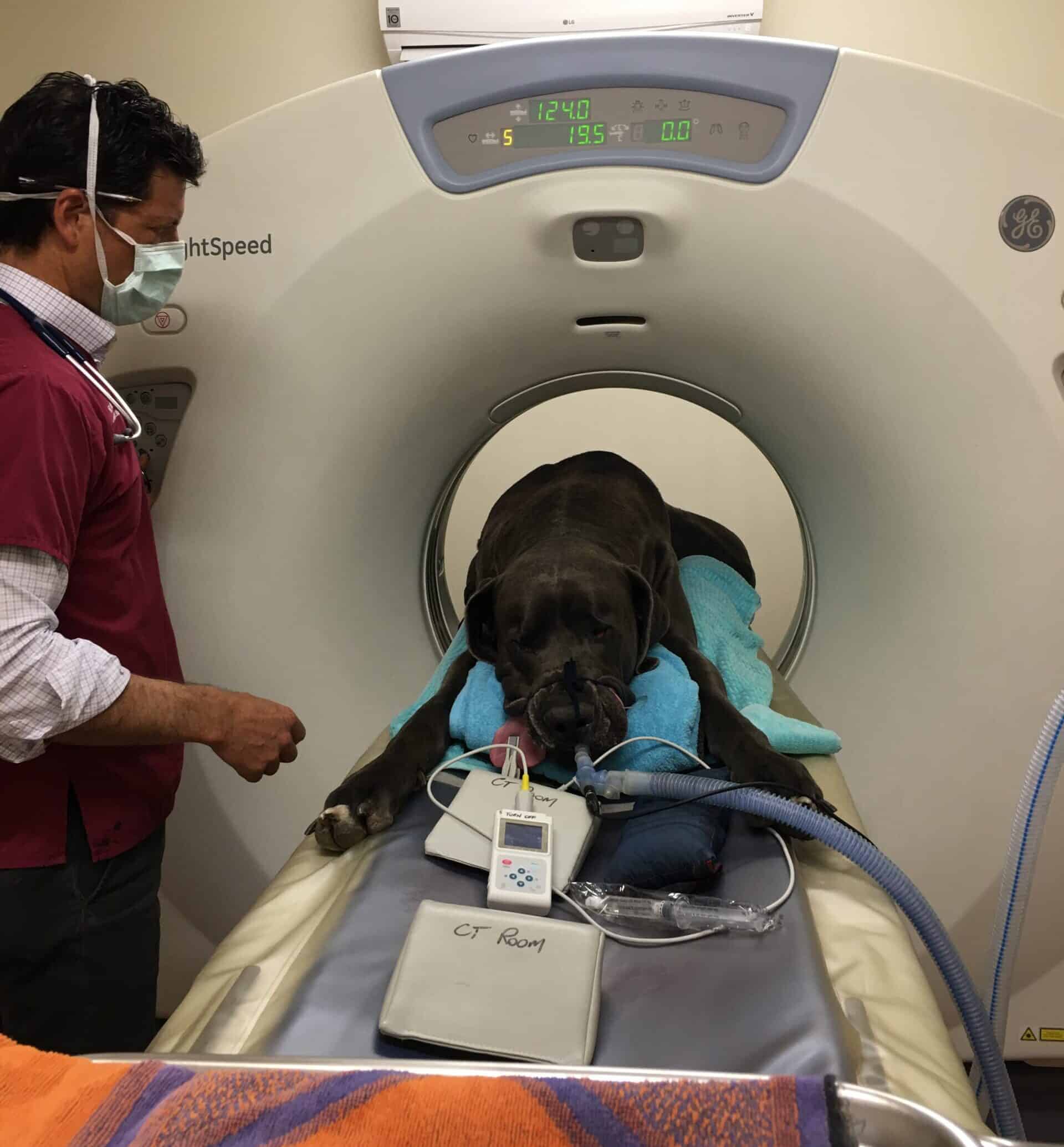
Dr Chris performing a CT scan on a Great Dane during Covid
Common Applications in Orthopedics
Fracture Evaluation: CT is particularly useful for assessing complicated fractures, such as those involving joints or when fracture fragments are small and difficult to see on X-rays.
Tumor Assessment: It helps in diagnosing and staging bone tumors, allowing veterinarians to evaluate the extent of the disease and plan treatment.
Joint Problems: CT can be used to examine joint surfaces and cartilage, aiding in the diagnosis of conditions like osteoarthritis or joint incongruity.
Pre-Surgical Planning: Detailed imaging helps surgeons plan complex orthopedic procedures by providing a clear understanding of the anatomical structures involved.
Considerations
- Anesthesia: Due to the need for precise positioning and stillness during the scan, anesthesia is often required for pets undergoing CT imaging.
- Cost: CT scans are typically more expensive than X-rays, which may influence the decision to use this modality.
- Radiation Exposure: While CT scans do involve radiation, the benefits often outweigh the risks when used appropriately.
Conclusion
CT scans are an invaluable asset in veterinary orthopedics, enhancing diagnostic capabilities and improving treatment outcomes. They are particularly beneficial for complex cases where detailed visualization is crucial. If you suspect your pet may need a CT scan, consult your veterinarian to discuss the best approach for diagnosis and treatment.
Fluroscopy
Intraoperative fluoroscopy is a real-time imaging technique used during surgical procedures, providing immediate visual feedback to surgeons. Here’s an overview of its application, benefits, and considerations in veterinary medicine, particularly in orthopedics.
What is Intraoperative Fluoroscopy?
Fluoroscopy involves the use of a continuous X-ray beam to create real-time images, allowing for dynamic visualization of internal structures. It is often used in conjunction with other imaging modalities, like X-rays and CT scans, to enhance surgical precision.
Applications in Veterinary Orthopedics
Fracture Fixation:
Fluoroscopy allows surgeons to visualize fracture alignment and hardware placement during orthopedic surgeries, such as the installation of plates or screws.
Joint Surgeries:
It aids in assessing joint stability and alignment during procedures like arthroscopy or joint reconstruction.
Minimally Invasive Techniques:
Fluoroscopy facilitates minimally invasive surgeries by allowing surgeons to navigate instruments and implants accurately while minimizing incisions.
Tumor Resection:
It can be used to visualize tumor margins during removal, ensuring complete excision while preserving surrounding healthy tissue.
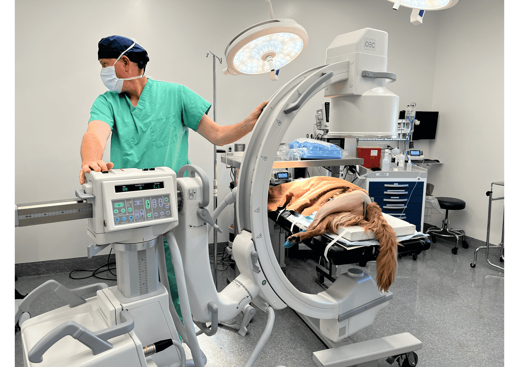
Positioning C-arm before hip replacement
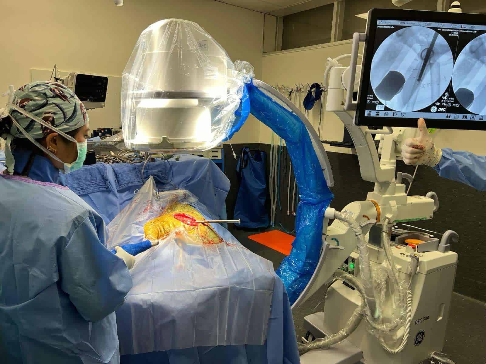
Use of C-arm with sterile plastic sleeve covers during hip replacement surgery in operating theatre to improve implant alignment
Benefits
Real-Time Feedback: Surgeons receive immediate imaging, allowing for adjustments during surgery to improve outcomes.
Enhanced Precision: Improves the accuracy of implant placement and alignment, which can lead to better recovery and function.
Reduced Surgical Time: Quick decision-making based on real-time images can help streamline procedures.
Considerations
Radiation Exposure: While fluoroscopy involves radiation, measures are taken to minimize exposure for both the patient and surgical team.
Cost: The use of fluoroscopy may increase the overall cost of the procedure due to equipment and operational requirements.
Technical Skill: Successful use of intraoperative fluoroscopy requires trained personnel to operate the equipment and interpret images effectively.
Conclusion
Intraoperative fluoroscopy is a powerful tool in veterinary orthopedic surgery, enhancing surgical precision and improving patient outcomes. It allows for real-time visualization of complex structures, making it invaluable for various procedures. If you have questions about its use in a specific surgery for your pet, consult your veterinarian or veterinary surgeon.
