IVDD / spinal
Types of IVDD
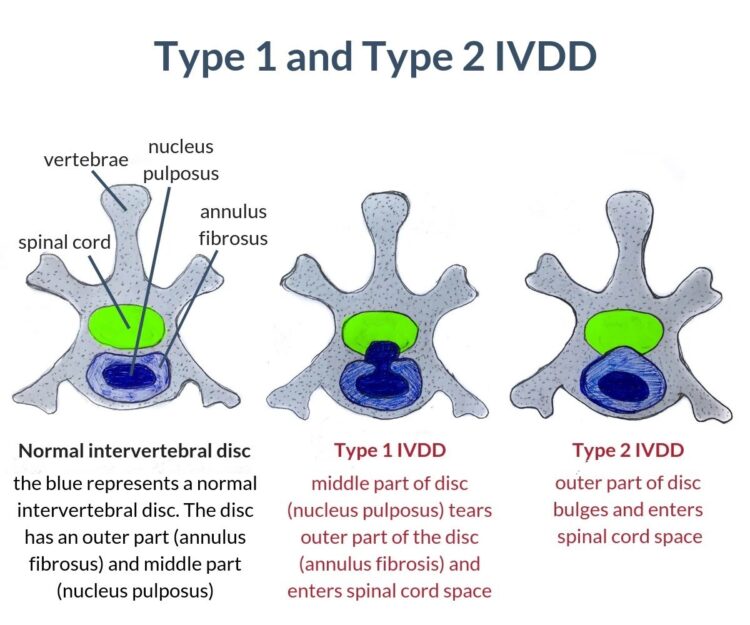
Type I IVDD:
- Description: This type is characterized by acute herniation of the disc material. The disc material bursts through the outer layer and compresses the spinal cord, often leading to sudden and severe symptoms.
- Common in: Chondrodystrophic breeds like Dachshunds, Cocker Spaniels, and Shih Tzus.
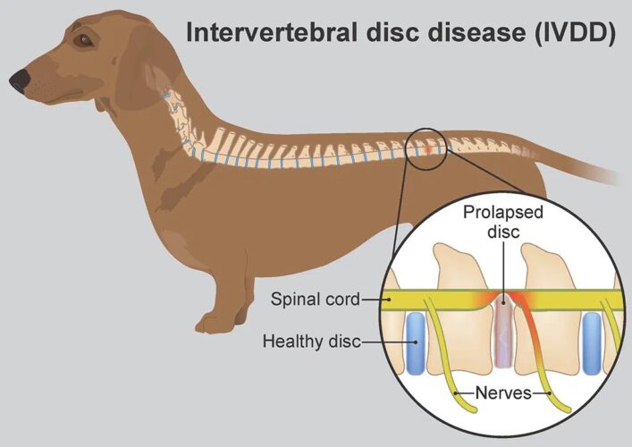
Type II IVDD:
- Description: This type involves a more gradual protrusion of the disc material, which causes chronic compression of the spinal cord. The disc bulges or herniates more slowly over time.
- Common in: Larger breeds like Doberman Pinschers and German Shepherds.
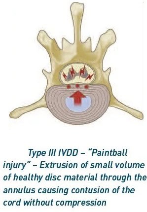
Type III IVDD:
- Description: This type involves acute disc extrusion where the disc material suddenly bursts into the spinal canal without significant compression, often causing severe pain and neurological symptoms.
- Common in: Can affect any breed, but less common than Type I and II.
Causes of IVDD
- Genetics: Certain breeds are genetically predisposed to IVDD, particularly chondrodystrophic breeds with abnormal disc degeneration.
- Age: Degenerative changes in the discs often occur with age, leading to reduced disc elasticity and increased risk of herniation.
- Injury or trauma: Sudden injury or trauma can exacerbate or lead to disc herniation.
Symptoms of IVDD
- Spinal pain: The dog may exhibit signs of pain, such as yelping when touched or reluctance to move. In severe cases, owners report spontaneous crying and screaming.
Severe neck pain
Severe back pain and pinched nerve
- Lameness: Difficulty walking or an unsteady gait, often with a tendency to drag the back legs.
- Weakness or paralysis: In severe cases, there may be partial or complete paralysis of the hind limbs.
- Loss of coordination: Difficulty with balance and coordination, particularly in the hind limbs.
Ridgeback with chronic progressive weakness and incoordination
- Reduced reflexes: Decreased or absent reflexes in the affected limbs.
- Incontinence: Loss of control over bladder or bowel functions in severe cases.
Neuro exam
Performing a neurological exam on a dog involves assessing various aspects of its nervous system. Here’s a step-by-step guide to help you conduct a thorough evaluation:
- Posture: Look for any abnormal postures (e.g., leaning to one side, head tilt).
- Gait: Observe the dog’s walking pattern. Look for limping, dragging, or uncoordinated movements.
This German Shepherd has weakness and incoordination
- Proprioception: Check the dog’s awareness of its body position by gently turning a paw over and observing if it corrects the position.
- Spinal pain: gentle pressure can be applied with incremental force to assess for the presence of a repeatable response.
Dachshund with loss of proprioception and focal spinal pain.
- Patellar Reflex: Tap the tendan below the kneecap to assess the amplitude of this reflex which tells us about the integrity of the spinal cord.
- Withdrawal Reflex: Pinch a paw and observe if the dog withdraws it quickly. This tests sciatic nerve function.
Assessing patellar tendon reflex using flexor
- Superficial Pain Perception: Gently pinch the toes to check if the dog reacts to pain.
- Deep Pain Perception: It is imperative to determine in severe cases that spinal cord is intact, as it greatly affects the overall chance of recovery for that patient. The most reliable way to assess pain perception is to apply focal pressure to each nail bed.
This paraplegic Bulldog was assessed for the presence of pain perception in the hind paws, to enable accurate grading and communication with the owner.
Neuro grades
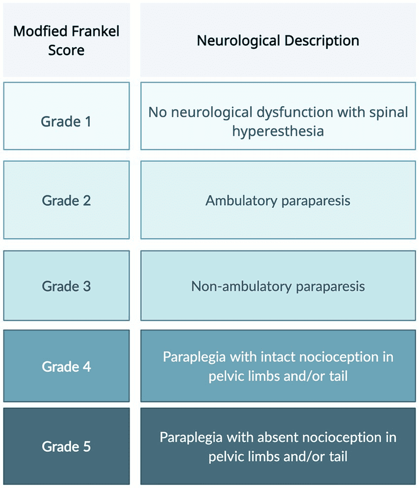
Explanation of terminology:
ambulatory = able to stand & walk unassisted
non-ambulatory = unable to stand unassistance
hyperesthesia = painful
paresis = reduced strength
plegia = no movement
para = back legs
tetra = front & back legs
Paraparesis: Dachshund with acute grade III IVDD = voluntary movement of the hind limbs
Tetraplegia: Poodle x with acute grade IV IVDD = no voluntary movement in all limbs
Diagnosis
- Physical examination: Veterinarians assess the dog’s gait, reflexes, and pain response to identify neurological deficits.
- Imaging: X-rays, MRI (Magnetic Resonance Imaging), or CT scans are used to visualize the spinal cord and discs, helping to confirm the diagnosis and determine the extent of the problem.
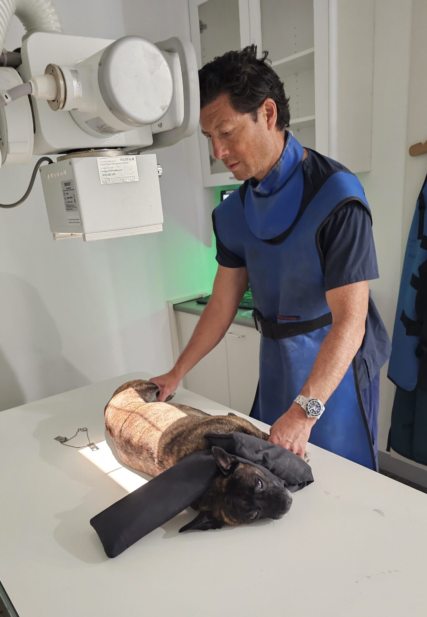
Dr Chris taking lateral spine xray
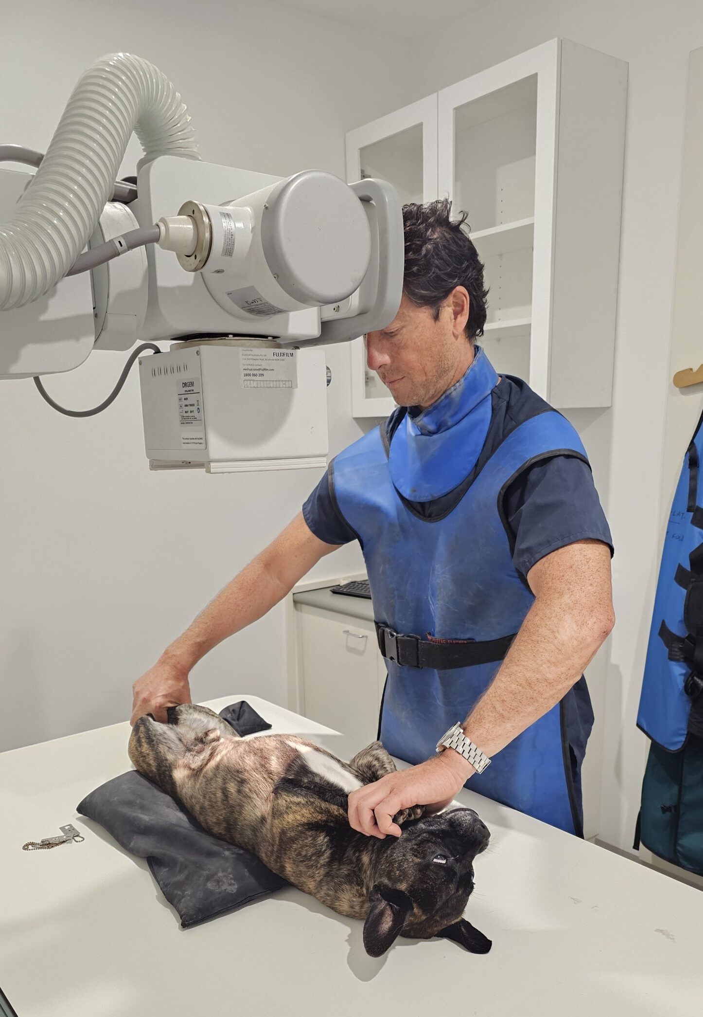
Dr Chris taking V/D xray
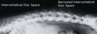
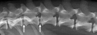

X-ray showing collapsed disc space

X-ray showing abnormal, mineralised disc

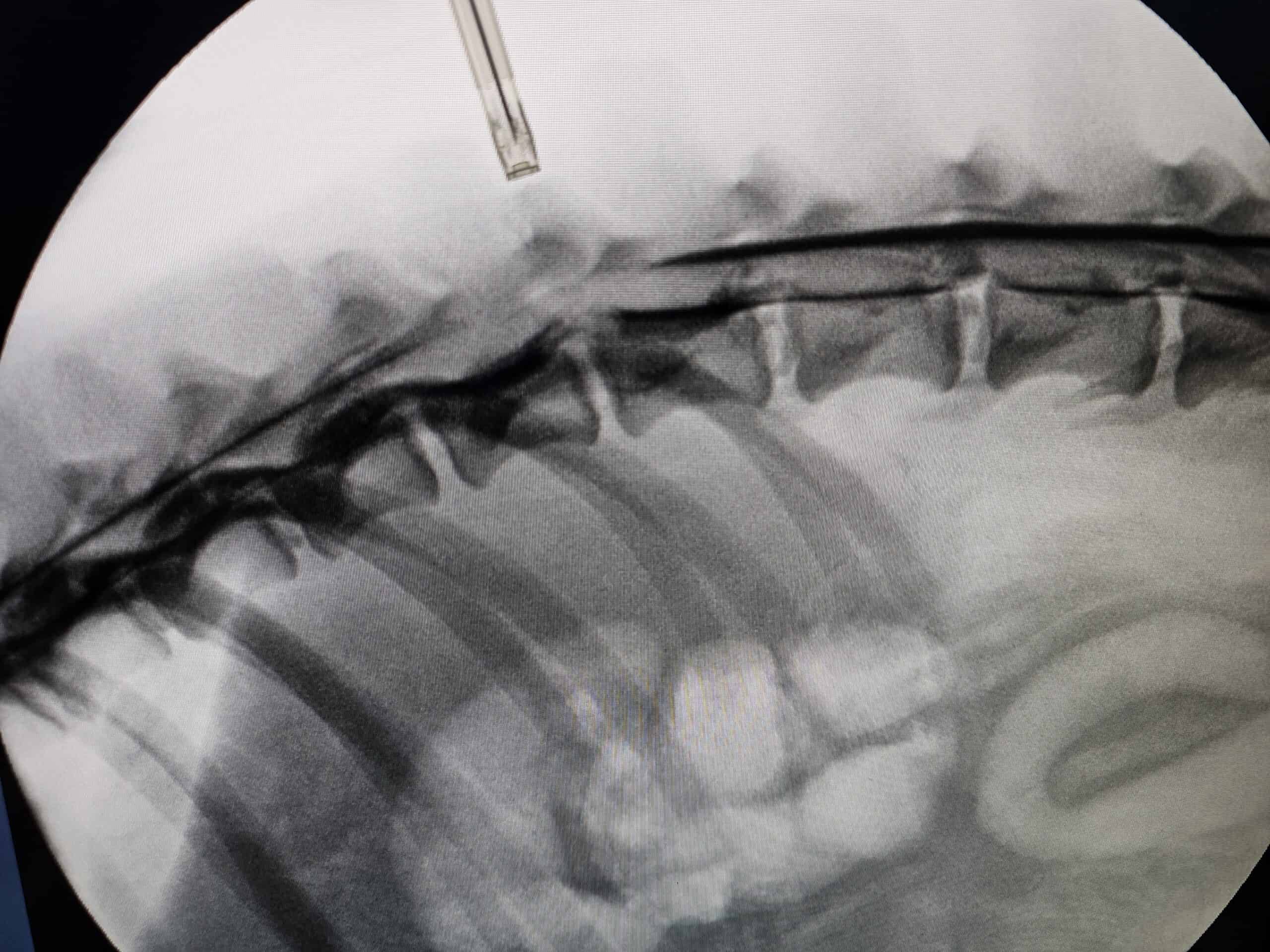
Fluoroscopy picture showing loss of contrast

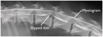
Contrast x-ray showing elevation of spinal cord
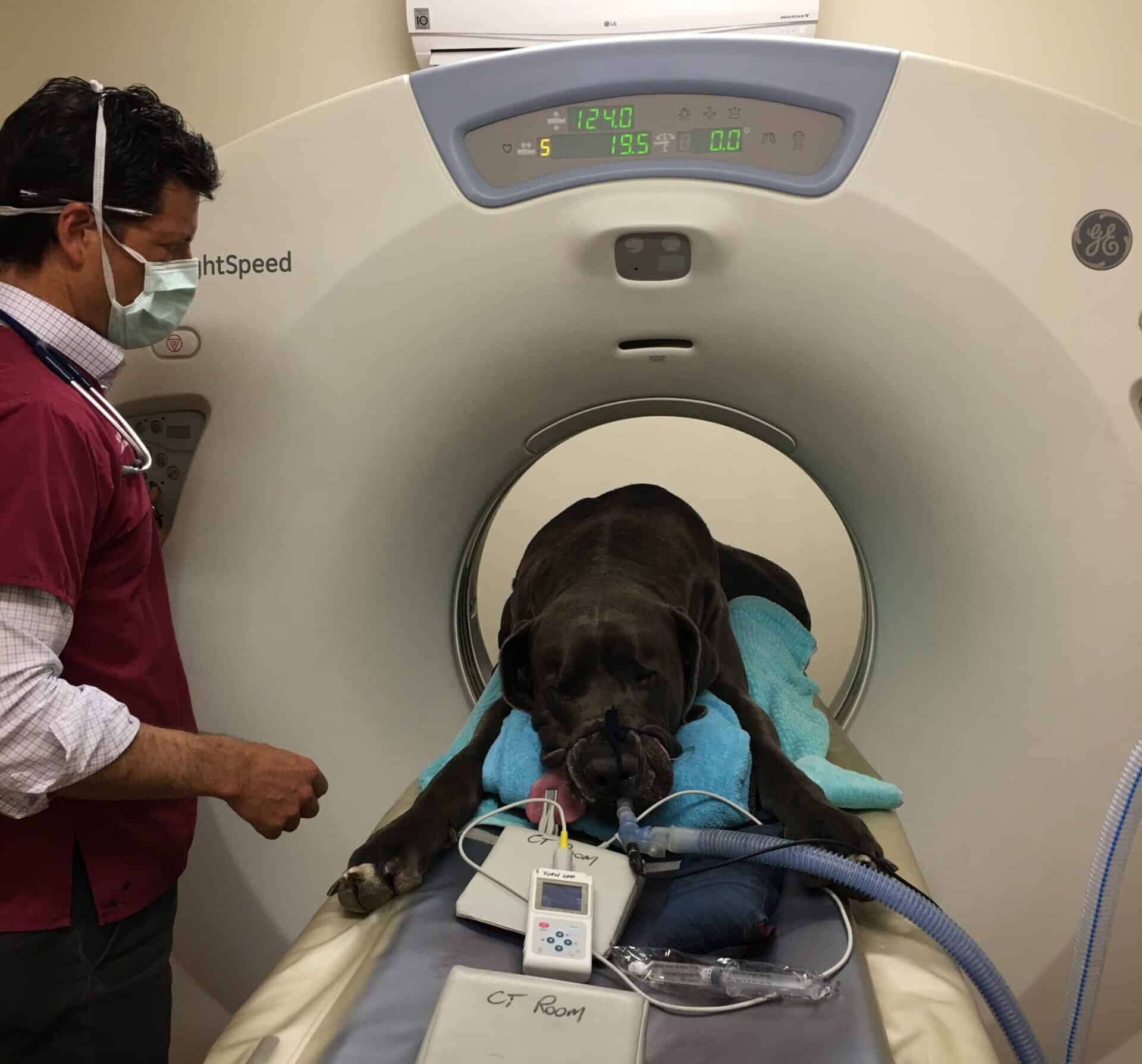
Dr Chris performing CT scan of spine

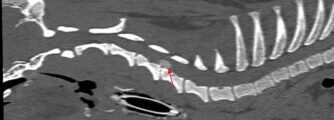
CT scan showing herniated disc in neck
CT scan showing herniated disk in back

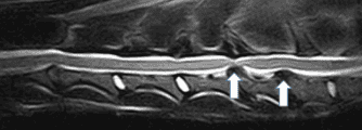
MRI scan showing two herniated discs in neck
Treatment options
Conservative management
- Medications: Pain relief and anti-inflammatory drugs (e.g., NSAIDs, steroids) to reduce pain and inflammation.
- Rest: Strict confinement and limited activity to allow the disc to heal and reduce stress on the spine.
- Physical therapy: Exercises and therapies to improve mobility and strength, often recommended after initial rest.
Surgical treatment
Surgery is often recommended for severe cases or when conservative management is ineffective. Common surgical procedures include:
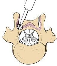
- Hemilaminectomy: Removal of part of the vertebra to relieve pressure on the spinal cord by removing the herniated disc material.
- Discectomy: Removal of the herniated disc material to relieve pressure on the spinal cord.
- Laminectomy: Removal of a portion of the vertebra to alleviate compression.
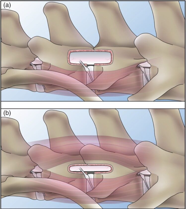
A = hemilaminectomy
B = pediculectomy
Case study: lumbar IVDD
Before surgery
7 days after surgery
Case study: neck IVDD
Before surgery
5 days after surgery
Post-surgical care
- Rehabilitation: Physical therapy to improve recovery, strength, and mobility.
- Restricted activity: Gradual reintroduction to activity, with careful monitoring to avoid complications.
- Follow-up visits: Regular check-ups with the veterinarian to assess recovery and ensure proper healing.
2 weeks postop Grade IV Spinal, recovering well
One week after spinal surgery in Dachshund
One week after spinal surgery in dachshund
Hydrotherapy
The buoyancy and resistance of water can be used to assist neurological patients in their recovery. An underwater treadmill allowed control of water depth and speed to encourage dogs to regain ambulant function faster.
Water level is lowered to increase demand on the pet. The water is warmed, filtered and chemically treated. Rehabilitation facilities can offer this service year round even in the cold Melbourne winter.
18 hours after neck spinal surgery called a “Ventral Slot”. Before surgery this dog had lowered head carriage and muscular spasms in neck.
Prognosis
- Mild cases: With prompt and appropriate treatment, many dogs recover well and return to normal or near-normal activity levels.
- Severe cases: Recovery may be more challenging, and some dogs may experience residual deficits or require long-term management. The prognosis often depends on the extent of spinal cord damage and the promptness of treatment.

7 days after neck (Type II IVDD) surgery. This Weimaraner was grade IV/V (tetraplegic) before surgery. Excellent long term prognosis.
IVDD can be a serious condition, but with early intervention and appropriate treatment, many dogs can experience significant improvements in their quality of life. If you suspect your dog may have IVDD, it’s crucial to seek veterinary care promptly to determine the best course of action.
MVO work closely with Casey Pet Emergency to offer immediate assessment and perform urgent spinal imaging and surgery.
