Patella luxation / MPL
Patella luxation, or kneecap dislocation, is a common orthopedic condition in dogs where the patella (kneecap) slips out of its normal position in the femoral groove. This condition can affect one or both knees and ranges from mild to severe, depending on how frequently and how far the kneecap dislocates. Here’s a detailed overview:
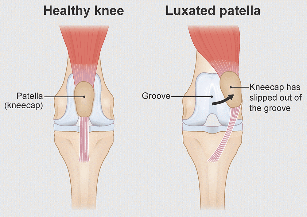
Causes of patellar luxation
- Genetic factors: Certain breeds are genetically predisposed to patellar luxation, especially small breeds like Pomeranians, Chihuahuas, Yorkshire Terriers, and Toy Poodles. However, larger breeds can also be affected.
- Congenital abnormalities: Abnormal bone structure, such as a shallow femoral groove or misalignment of the leg bones, can predispose a dog to patellar luxation.
- Trauma: Injury to the knee can cause or exacerbate patellar luxation, especially if it leads to damage of the ligaments or joint capsule.
Grades of patellar luxation
Patellar luxation is categorized into four grades, which help determine the severity of the condition and the appropriate treatment:
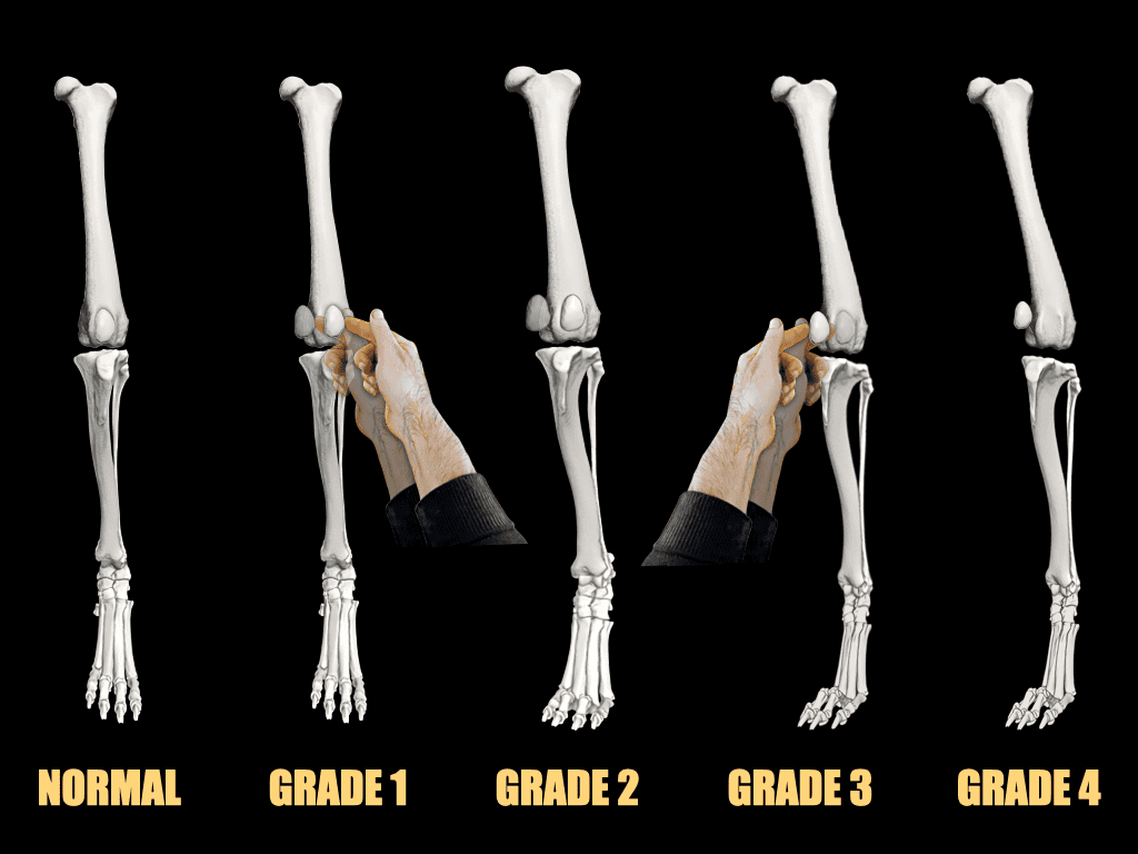
Grade I
- Description: The patella can be manually luxated but returns to its normal position on its own.
- Symptoms: Minimal, often asymptomatic; may occasionally cause a “skipping” gait.
Grade II
- Description: The patella luxates spontaneously or with manual manipulation but returns to its normal position on its own.
- Symptoms: Intermittent lameness, occasional skipping or hopping.
Grade III
- Description: The patella is luxated most of the time but can be manually replaced.
- Symptoms: Persistent lameness, abnormal gait, discomfort.
Grade IV
- Description: The patella is permanently luxated and cannot be manually repositioned.
- Symptoms: Severe lameness, abnormal limb structure (bow-legged or knock-kneed appearance), significant discomfort.
Symptoms of patellar luxation
- Lameness: Intermittent or persistent lameness, often more pronounced after exercise.
- Skipping gait: Dogs may “skip” or hop on one leg, especially when the kneecap is out of place.
- Abnormal posture: Bow-legged or knock-kneed stance, particularly in more severe cases.
- Pain: Discomfort or pain when the kneecap luxates, though mild cases may not show obvious signs of pain.
- Reluctance to move: Some dogs may be reluctant to walk, run, or jump, especially if the condition is severe.
Diagnosis
- Physical examination: Veterinarians can often diagnose patellar luxation through a physical examination, manipulating the knee to see if the patella moves out of place.
MPL exam standing
MPL exam recumbant
- X-Rays: Imaging helps assess the severity of the condition, evaluate the bone structure, and determine the extent of any secondary arthritis.
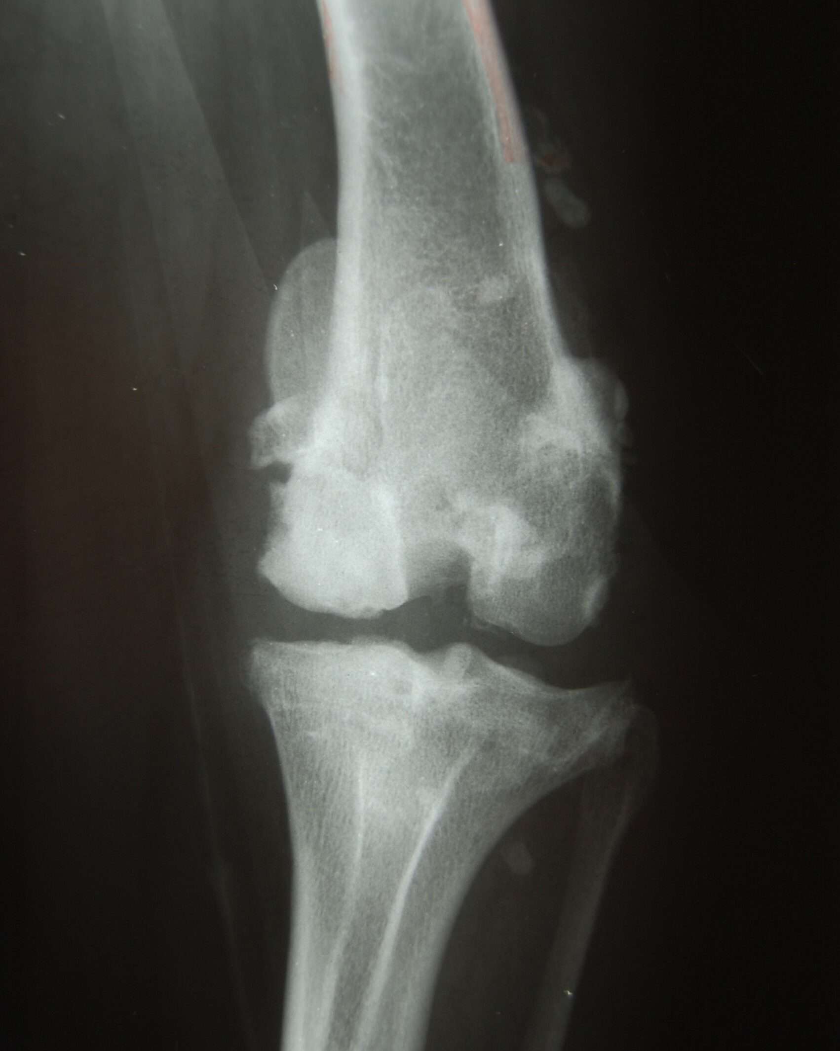

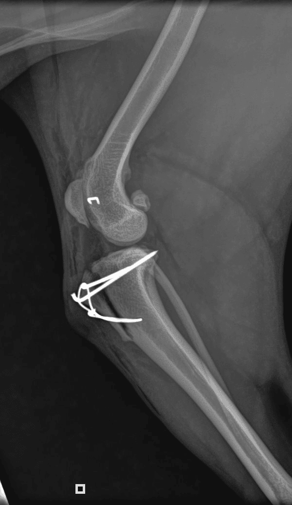
Typical postoperative x-rays.
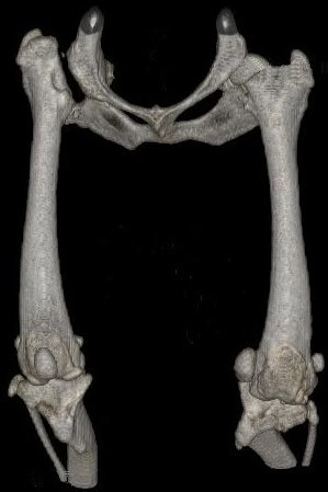
- CT scan: three dimentional imaging of the skeleton is now available at specialist centers. This enables surgeons to comprehensively plan corrective surgery. 3D printing technology allows bone models to be manufactured, allowing a rehearsal surgery to be performed.
Treatment options
Non-surgical treatment
- Weight management: Keeping the dog at a healthy weight reduces stress on the knees.
- Physical therapy: Strengthening the muscles around the knee can help stabilize the joint.
- Pain management: Non-steroidal anti-inflammatory drugs (NSAIDs) and other pain relief medications can help manage discomfort.
- Activity modification: Reducing high-impact activities to prevent exacerbating the condition.
Surgical treatment
Surgery is typically recommended for grades II-IV or when conservative treatments are not effective:
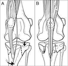
- Tibial Tuberosity Transposition (TTT): The attachment point of the patellar tendon is moved to a more central position on the tibia, helping to realign the patella.
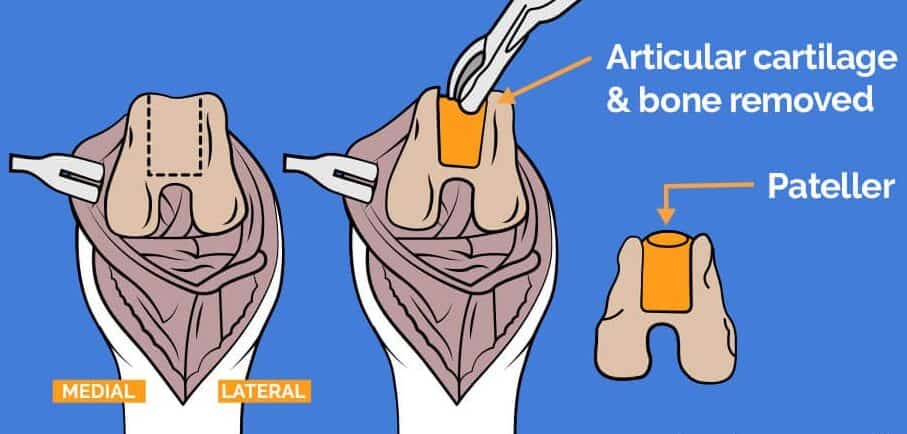
- Trochleoplasty: The femoral groove is deepened to better hold the patella in place.
- Lateral imbrication: Tightening the tissues on the outside of the knee to prevent the patella from slipping out of place.
- Medial release: Loosening tight tissues on the inside of the knee, which may be pulling the patella out of its normal position.
Post-surgical care
- Restricted activity: Strict rest and limited movement are essential during the initial healing period to ensure the surgical repair is successful.
- Physical therapy: Post-operative rehabilitation is often recommended to restore strength and flexibility in the knee.
- Pain management: Medications to control pain and inflammation during recovery.
- Follow-up visits: Regular veterinary check-ups to monitor healing and ensure proper recovery.
Prognosis
- Mild cases (Grades I and II): Dogs with mild patellar luxation often manage well with conservative treatment and may not require surgery.
- Moderate to severe cases (Grades III and IV): Surgery generally has a good success rate, with most dogs returning to normal or near-normal activity levels after recovery.
A litter of Basenji pups all affected with grade IV MPL
Prevention
- Selective breeding: Avoid breeding dogs with a known history of patellar luxation to reduce the risk in future generations.
- Maintaining a healthy weight: Keeping dogs at an ideal weight reduces stress on the joints and can help prevent or manage patellar luxation.
- Regular exercise: Maintaining good muscle tone around the knee can help stabilize the joint.
Patellar luxation is a manageable condition, and with proper care, dogs can lead comfortable, active lives. If you suspect your dog has patellar luxation, consult your veterinarian for an accurate diagnosis and appropriate treatment plan.
